05 Jul Motor Center Structure and Function
The past few posts in this section have focused on the Cerebrum where thought seems to be centered: we may compare this to a computer. The Cerebellum is where signals for the muscles’ motor control originate: we may compare this to a robot. My point in looking at this is to reinforce the heterogeneity perspective (i.e. that traditional neural networks do not look like the brain), and to support my feeling that thought and action are intrinsically tied at the core of context.
 Layers in the Cerebellum Structure
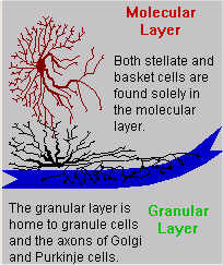
Today, I will focus on the cerebellum. This is the part of the brain that controls our muscle movement. I will treat it almost like a separate organ today. In this illustration, the red, blue and bright green text colors are used to represent layers. Blue also represents a cell type because one layer consists solely of that type of neurons (Purkinje Cells).
The molecular layer is the outermost layer of the cerebellar gray matter. It contains the dendrite processes of all five types of cells. Stellate cell bodies and axon(s) are only found in the molecular layer. Basket cell bodies straddle the Purkinje layer – their axons penetrate both the Purkinje layer and the molecular layer. Purkinje cells send their dendrite processes (branches) through the molecular layer, their somas are only in the Purkinje layer, and their axons protrude through the granular layer and into the white matter.
Whereas the structure of the cerebrum was fairly rigidly determined, the cerebellum is more stratified. Purkinje cell axons constitute the only source of output from the cerebellum [Kuffler, et al.,1984, p. 11]. They also have direct input links through their extensive dendritic arborization. Thus, though there are many other cells of different types that participate in the processing functions of the cerebellum, each Purkinje cell is involved more extensively than any of the others in both input and output.
| Understanding Context Cross-Reference |
|---|
| Click on these Links to other posts and glossary/bibliography references |
|
|
|
| Prior Post | Next Post |
| Centers of Attention and Consciousness | Understanding Subtext |
| Definitions | References |
| arborization | Kuffler 1984 |
| Purkinje Cells | Purkinje World |
 Stellate & Basket Cells
Stellate cells in the cerebellum are pictured in the illustration. They are located entirely in the molecular layer. Both the afferent and efferent fibers form synapses with other cells and fibers in the molecular layer. This localization may point to a function that could be ascribed to the layer itself. It might also be ascribed to cell fibers acting within the layer, including Purkinje and Golgi dendrites.
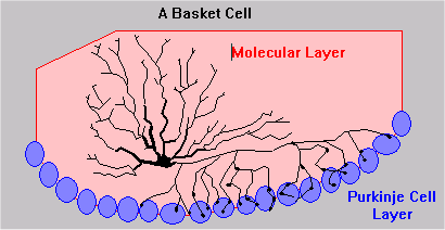
Basket cells have substantial dendrite branching or arborization. They usually have a single primary axon with termini leaving the axon to form synapses all along its length. Because these synapses are in the Purkinje cell layer that consists primarily of Purkinje cell bodies or somas, it is reasonable to assert that most of the basket cell outputs go directly into the cell bodies of Purkinje cells.
Purkinje cells have substantially greater dendrite arborization than any of the other cells. Their axons normally plunge all the way to the  white matter, often sending secondary terminals back through the Purkinje layer. This morphology differs profoundly from that of Golgi and granule cells, both of which have multiple axon processes.
Both stellate and basket cells are found solely in the molecular layer. The granular layer is home to granule cells and the axons of Golgi and Purkinje cells.
Granule, Golgi Basket and Granule Cells
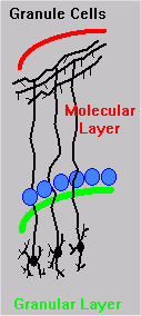
 The bodies of granule cells are in the granular layer. Their dendrites extend radially within the granular layer. Their axons extend well into the molecular layer. These axons send out long parallel fibers horizontally to propagate input to the Purkinje cells’ dendrites and those of cells found only in the molecular layer. The dendrites may have termini in close proximity to the soma, or they may extend into the white matter. Mossy fibers carry impulses from other areas of the brain to granule cells in the cerebellum. Golgi cell axons provide feedback input to granule cell dendrites.
Golgi cell bodies are also in the granular layer. Their dendrites extend into the molecular layer. Dendrite arborization appears more regular than that of stellate and basket cell processes. The most distinguishing trait of Golgi cells is that the axons also display complex arborization, though it is limited to the granular layer. Where Purkinje cells usually have a single axon extending deep into the white matter and other areas of the brain, Golgi cells have multiple feedback outputs close to the cell body that display a more regular symmetry.
In the cerebellum, granule cell bodies are in the granular layer while their axons reach through the molecular layer to send parallel fibers branching near the surface of the cortex. The dendrites are fairly short.
Basket cells in the cerebellum straddle the Purkinje cell layer with dendrites receiving impulses throughout the molecular layer and multiple axons sending inhibitory impulses to Purkinje cells.
 Cerebellum Function
The functional diagram at right shows the information flow in the nervous system that regulates both voluntary and involuntary activity. The five senses give us cues we may act on. In the cerebral cortex and subcortical areas we interpret cues, choose courses of action, and adjust as circumstances dictate. The direct link between receptors and the muscles permits rapid reflexes. For deliberate actions, however, information passes through the coordinating centers first. Click on the diagram for a look at the cerebellum circuitry.
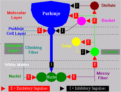
Cerebellum Circuit
The circuitry of the cerebellum, like that of the cerebrum, is very specific. When we try to imitate this circuitry in computer hardware and/or software, we cannot assume that the flow of impulses is totally random or the same for both positive and negative impulses. The cerebellum coordinates movements of muscles throughout the body to support dexterous and precise movements and the proper application of force to achieve the objectives of planned motions. Sensory input/feedback to the cerebellum helps coordinate muscle activity.
Structural and Functional Complexity
The interplay of structures that support functions, both at the cellular level and at the organ level, and the complexity of their interactions constitute the many dimensions of brain processes. Automating these tasks on computers demands a multi-dimensional approach. More on this to come…
| Click below to look in each Understanding Context section |
|---|


