29 Aug Layers of Brain Complexity

Hierarchy: A Structural Aspect of Thought
The complexity of the cerebrum is necessitated by its function as the center of cognition. Before looking at cerebral structure, consider some of the functions of thought. Thinking involves accessing memory to find what perceived data is stored, what other data in memory it is associated with, and what is the nature of associations. For new items, thinking often involves conscious attempts to classify things into superclasses and analyze them into their constituent parts. This involves search procedures through a person’s body of knowledge and reasoning about classes of associations.
In this blog, I’m looking closely at these connections between body and mind, between cognition and communication, because I believe that this has been lacking in some artificial intelligence or cybernetics research in the past. I have a hard time accepting views of thinking and communicating that exclude any interconnected domains of knowledge, and want to look in sufficient detail at all the factors that could contribute to our ability to understand one another.
| Understanding Context Cross-Reference |
|---|
| Click on these Links to other posts and glossary/bibliography references |
|
|
|
| Prior Post | Next Post |
| Brain Correlation Processes | Sensory Input to the Brain |
| Definitions | References |
| cerebrum input functions | Kuffler 1984 |
| memory cognition | Berne and Levy Cortex |
| classify data associations | Scott 1995 |
The simple diagram below shows one way people deal with knowledge: we classify it into hierarchies. A simple example of hierarchical classification is the way biologists have categories for all living things. At the top are the kingdoms of plants and animals. Organisms are further categorized by phylum, genus, and species. Though little is known about how the brain makes hierarchical classifications, a description of cerebral structure can assist in analyzing how this structure may be used to accomplish tasks such as hierarchical categorization.
Cerebrum Layers
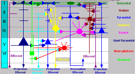
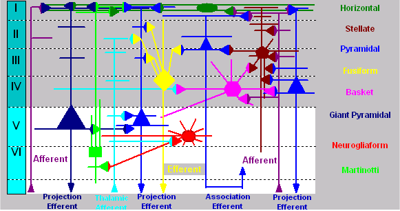
There are six layers identified in 90% of the cerebrum. The chart below briefly describes them.
| # | Name | Depth | Cell body types | Fiber patterns |
| I | Molecular | 200 µm | horizontal – stellate Tangential layer; (plexiform) or granule | horizontal fibers |
| II | External | 200 µm | pyramidal -granule Dysfibrous layer granular Martinotti | shorter fibers |
| III | External | 450 µm | small pyramidal-Suprastriate layer pyramidal granule – Martinotti | mostly horizontal |
| IV | Internal | 200 µm | pyramidal -External band of granular – granule – Martinotti | Baillarger (h+v) |
| V | Ganglionic | 250 µm | giant pyramidal Interstriate layer (internal granule -Martinotti horizontal with pyramidal) | more vertical |
| VI | Multiform | 250 µm | fusiform – pyramidal Infrastriate layer (Fusiform) granule -Martinotti | vertical/horizontal |
The area shown below layer VI is Sub-cortical white matter with myelinated fibers but no cell bodies . The structural morphology of cerebral cells, their organization, and their connections suggest that their structures and functions are equally diverse. The range of connectivity in cerebral cells is from dozens of synapses in small cells to tens of thousands in the largest. Pyramidal cells are the most numerous in the cortex. Small pyramidal cells can be 20-30 micrometers while giant pyramidal cells (the minority) can be from 70 to over 100 micrometers. Giant pyramidal cells (Betz cells) are characteristic of the motor area (4) in layer V.


Cell Shapes and Links
Links between neurons are formed at synapses. These will be discussed in greater detail in Sections 2 and 3. For now, take a look at the illustration at right. It shows the three general types of incoming synapses. Outgoing synapses can be any of the three types, but because they occur less frequently, they are all the same color on the graphic. Note that input links occur anywhere on the cell membrane, with axodendritic being the most common by far. Differences between cell morphologies and locations in the brain are usually attributed to functional specialization. Some of the cells may serve different roles, requiring limited links with certain other specialized cells. The types of links may also be different: some favor axodendritic while others prefer axosomatic or axoaxonic. Purkinje cells are known to provide the output channel, suggesting intriguing possibilities for processing and coordinating roles for the other cell types.
The cerebrum is far more complex than the cerebellum. This could be due to the complexity of functions of the cerebrum, which range from motor control to symbolic communicative processing to reasoning. These functions will be discussed more fully in later posts. For now, the foundational point for cybernetic modeling is that a simple model will not sufficiently represent this much brain complexity. A highly distributed model with large numbers of very simple processing elements is also unlikely to be able to replicate the processes handled by such a complex system.
| Click below to look in each Understanding Context section |
|---|


